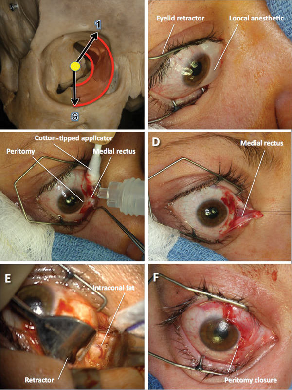
Fig. 3.

Medial micro-orbitotomy. (A) This approach gives access to lesions located anterior and medial in the orbit. (B) An eyelid retractor is placed and local anesthetic injected where the peritomy will be performed. (C) After the conjunctiva is incised around the cornea and relaxing conjunctival incisions are made, the medial rectus muscle is detached and (D) retracted medially with a suture. (E) The eye globe is retracted laterally and the intraconal fat exposed. (F) After the lesion has been excised, the medial rectus muscle is reattached at its insertion site on the globe with a 6-0 absorbable suture, and the conjunctiva is closed with interrupted sutures.
