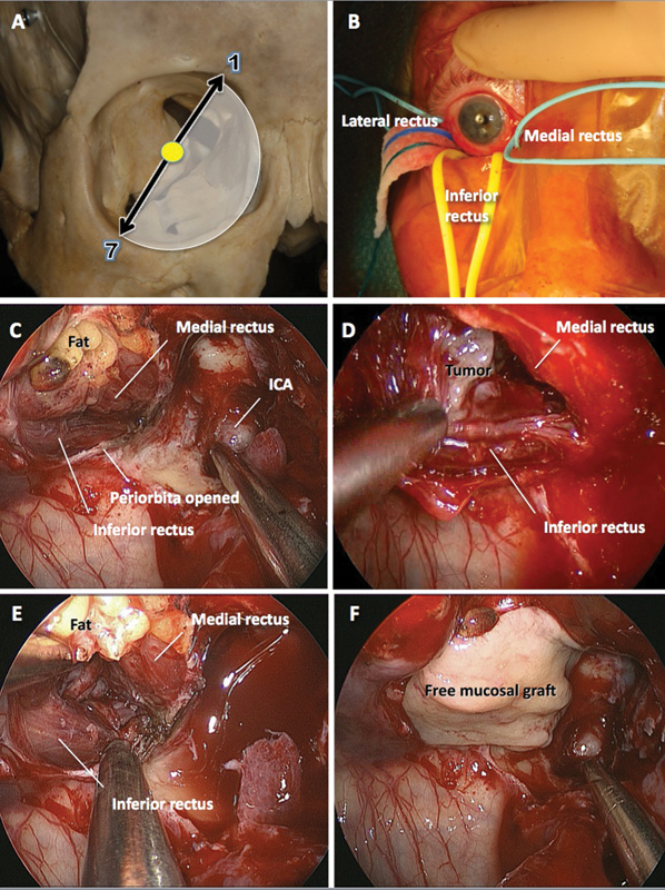
Fig. 4.

Endoscopic endonasal approach. (A) Clock model showing the extent of the orbit that can be exposed through this approach. (B) The insertions of the rectus muscles to the globe are identified and controlled with vessel loops. (C) Endoscopic view of the medial aspect of the orbital apex after a portion of the periorbita has been excised. The internal carotid artery (ICA) is visible medially. The window between medial and inferior rectus muscles is “closed.” (D) After external retraction on the medial and inferior rectus muscles by pulling the respective vessel loops, the surgical window in now “open” and the tumor is identified and (E) excised. (F) The periorbital defect is covered with a free mucosal graft harvested from the removed ipsilateral middle turbinate.
