
Fig. 1.
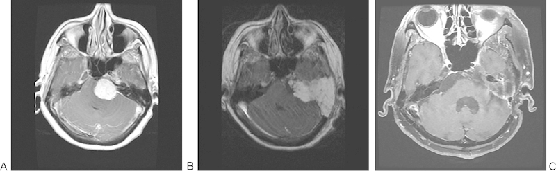
Patient with a 3.5-cm left petroclival meningioma excised by an extended translabyrinthine approach (subtotal resection as tumor capsule left on brainstem). (A) Preoperative T1-weighted magnetic resonance imaging (MRI) with gadolinium. (B) Immediate postoperative T1-weighted MRI with gadolinium. (C) A 5-year postoperative fat-saturated T1-weighted MRI with gadolinium.