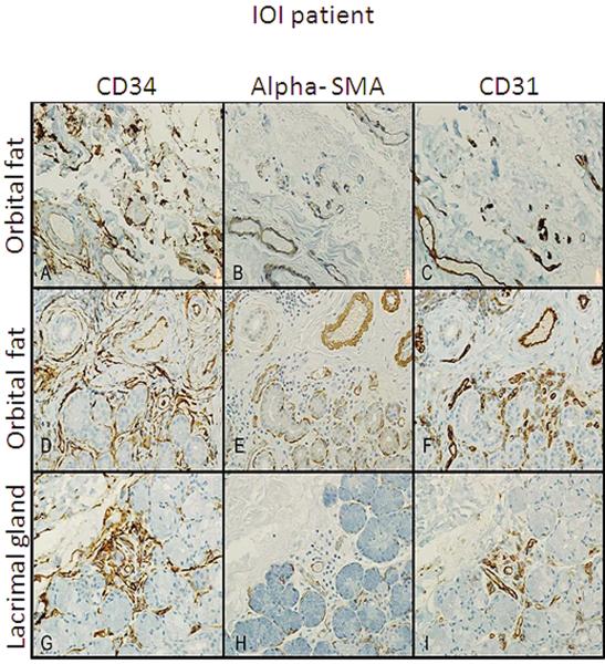
Figure 1.
Immunohistochemistry of orbital fat from three IOI patients stained with CD34 alpha SMA and CD31. Spindle-shaped CD34+ cells (A, D,and G), are present in a perivascular distribution with early fibrosis and mild chronic inflammation. α–SMA+ cells (B, E, and H) showed staining of only smooth muscle of blood vessels and myoepithelial cells in sequential tissue sections. CD31+ cell staining was limited to blood vessel endothelial cells(C, F, and I). All 400X of the same region.

