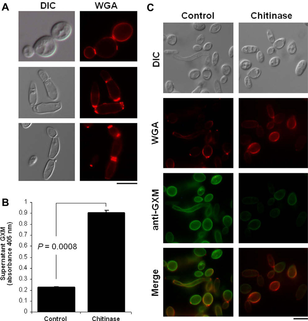
Fig. 5.
GXM anchoring to the cell wall of T. asahii (isolate CBS2479) involves chitin oligomers. (A) Incubation of T. asahii cells with the lectin WGA reveals that external chitin-like structures are concentrated in cell division sites. Similar results of three different experiments prepared under the same conditions are shown. (B) ELISA of supernatants of T. asahii after incubation in PBS (control) or in the same buffer supplemented with chitinase reveals that GXM is released after exposure to the enzyme. (C) Incubation in the presence of the enzyme also changes the pattern of WGA binding to T. asahii. Analysis of the fungal cells after the conditions described in B confirms the reduction in the content of surface GXM after chitinase treatment. Fungal cells observed under differential interferential contrast (DIC) and fluorescence mode (anti-GXM and WGA) are shown. Scale bars, 3 µm.

