Figure 3.
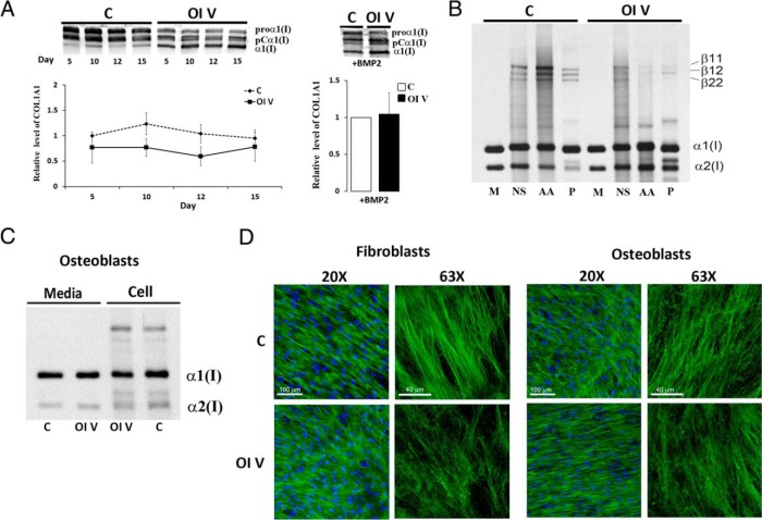
Type I collagen secretion and deposition. A, Left panel, Representative Western blot of type I procollagen secretion from control (C) or patient 3 (OI V) osteoblasts (OBs) stimulated with osteogenic media for up to 15 days, or, right panel, for 10 days with BMP2 treatment. Patient 3 has decreased collagen secretion and was equal with BMP2 treatment. B, Extracellular collagen matrix deposited by control (C) or patient 3 (OI V) OBs. Normalized densitometry of control and OI V α1(I) chains after balanced loading revealed a 3.5-fold decrease in mature cross-linked collagen as well as a decrease in cross-linked β-forms in acetic acid (AA) and pepsin (P) fractions. C, Steady-state type I collagen protein in osteoblasts from control (C) and patient 3 (OI V). Migration of the α1(I) and α2(I) chains is normal in the patient. D, Immunofluorescence microscopy of collagen matrix deposited in culture by control (C) and patient 3 (OI V) fibroblasts and osteoblasts. Type V OI matrix has patchy, parallel fibers in comparison with the networked, well-formed fibers in control. M, media; NS, neutral salt.

