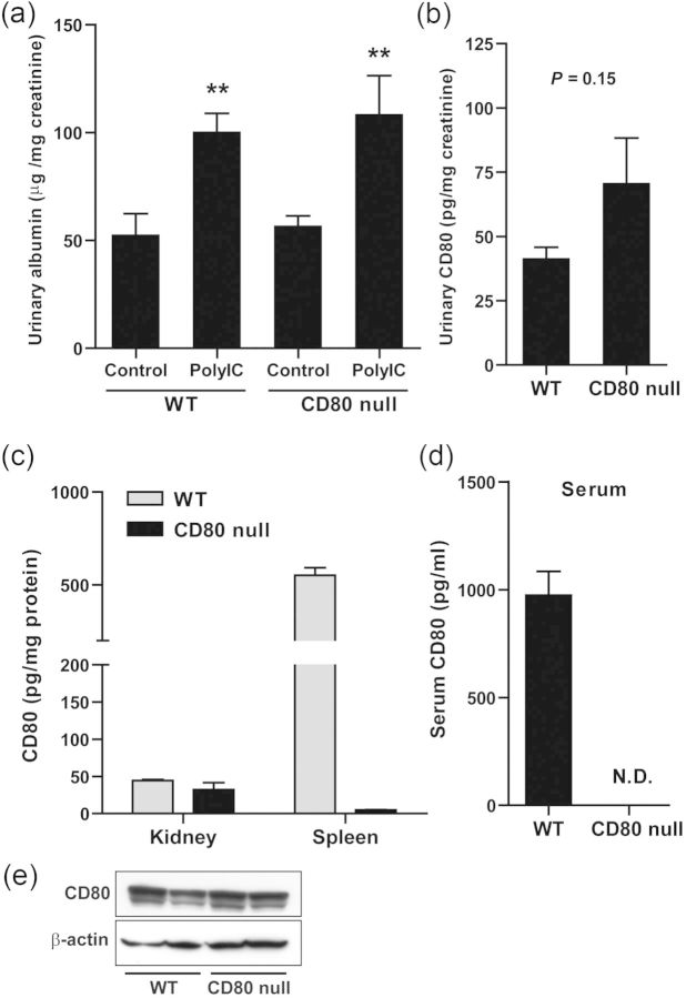
FIGURE 6:
The examinations using CD80 null mice. (a and b) WT mice and CD80 null mice were intravenously injected with polyIC-LMW. (a) Twenty-four-hour urine was collected at 24 h (0–24 h). Control urine was collected 1 week prior to the injection of polyIC-LMW. Urinary albumin-to-creatinine ratio was measured. The results represent means ± SEM (n = 6). **P < 0.01 versus respective control. (b) Urinary concentration of CD80 protein was determined by ELISA, and was corrected by creatinine concentration. The results represent the means ± SEM (n = 5). (c) CD80 contents in the kidney and spleen of WT mice and CD80 null mice measured by ELISA. The results represent means ± SD (n = 3). (d) Serum CD80 concentration measured by ELISA. The results represent the means ± SD (n = 3). (e) Representative images of western blotting for CD80 using the kidney of WT mice and CD80 null mice.

