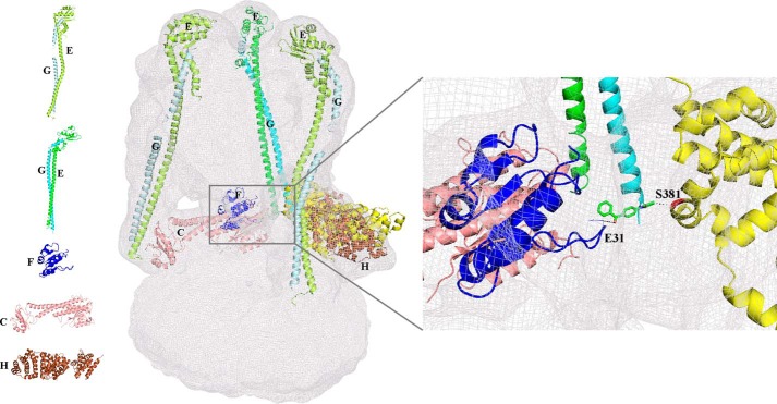
FIGURE 1.
Arrangement of the existing individual atomic subunit structures in the EM map of the S. cerevisiae V-ATPase. Subunits C (1U7L; salmon), H (1HO8, brown), and F(1–94) (4IX9, blue) from S. cerevisiae were fitted into the EM map. The two conformations of EG subunits, the straight (4DL0; green and cyan) and more bent (4EFA; lemon and pale cyan) are fitted to the three peripheral stalks. Inset, region of the EM map showing the interaction of modeled subunit H (Ser-381) (yellow) through the sulfhydryl cross-linker 4-(N-maleimido)benzophenone (62) (stick; green) to the S. cerevisiae subunit F(1–94) (Glu-31). Left panel, schematic representation of the structures of the individual S. cerevisiae subunits C (1U7L; salmon), F(1–94) (4IX9, blue), H (1HO8, brown), and EG in two conformations, straight (4DL0; green and cyan) and bent (4EFA; lemon and pale cyan).

