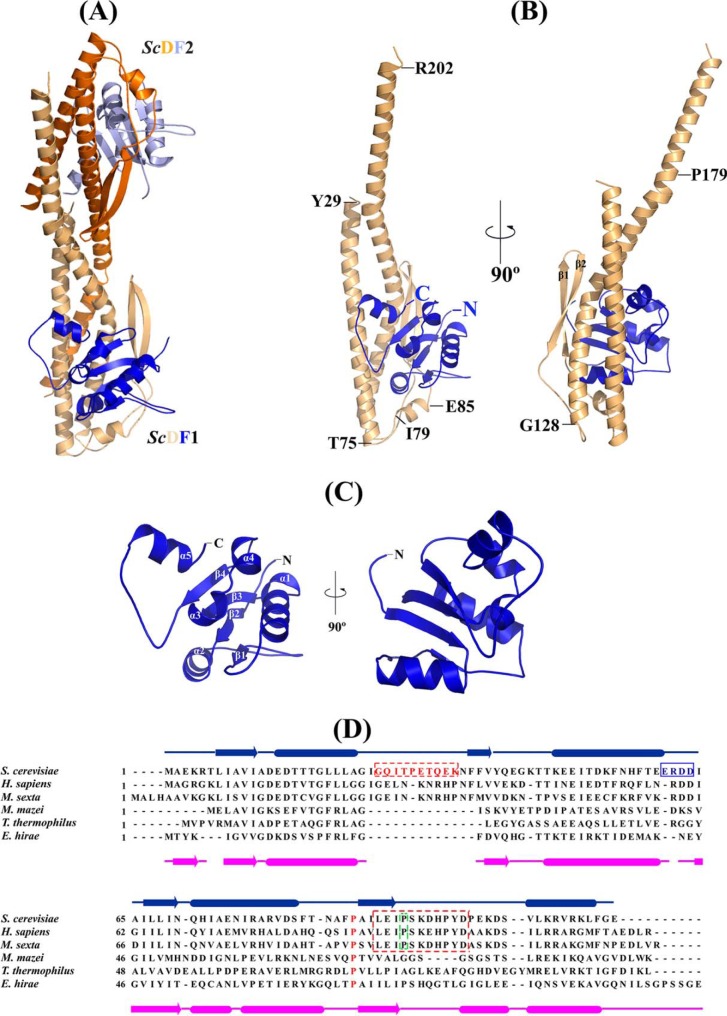
FIGURE 2.
Crystal structure of S. cerevisiae subunit DF complex. A, crystal structure of DF complex of the S. cerevisiae V-ATPase in an asymmetric unit; ScDF1 is shown in wheat and blue, and ScDF2 is shown in orange and light blue, respectively. B, schematic representation of the ScDF1 complex; ScD and ScF are shown in wheat and dark blue, respectively. Proline 179, which is conserved in all the eukaryotic ATP synthases, is labeled. C, schematic representation of ScF1 (dark blue), with the unique C terminus helix conformation and the α-helices and β-strands labeled. D, sequence alignment of subunit F from different ATPases. The secondary structure elements of subunit F from E. hirae and S. cerevisiae are shown. The conserved sequence in the eukaryotic ATP synthase is shown within the box. The 26GQITPETQEK35 loop of the F subunit is highlighted in red. The conserved Pro-89 is highlighted in red and is present in V-ATPases as well as A-ATP synthases. Proline 95, which is conserved in eukaryotic V ATPases, is highlighted by a green frame.

