FIGURE 3.
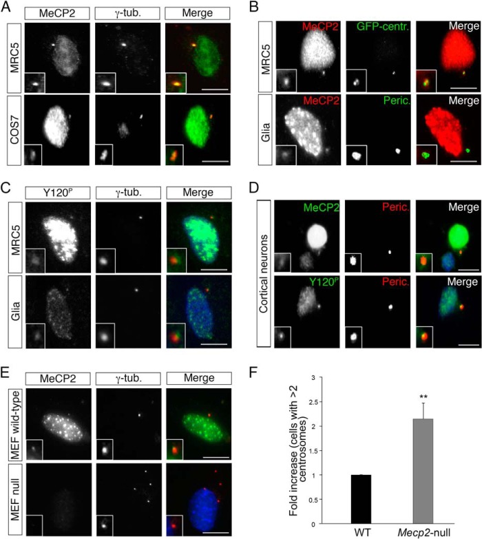
MeCP2 is targeted to the centrosome in different cell lines. A, MRC-5 and COS-7 cells were immunostained with anti-MeCP2 (green) and anti-γ-tubulin (γ-tub, red) antibodies (n > 3). B, MRC-5 cells (top panels) expressing an exogenous GFP-centrin (GFP-centr.) fusion protein (green) were stained with anti-MeCP2 (red). Mouse glial cells (bottom panels) were costained with anti-MeCP2 (red) and anti-pericentrin (Peric., green) antibodies (n = 3). C, MRC-5 and mouse glial cells were stained with anti-Tyr(P)-120 (green) and anti-γ-tubulin (red) antibodies (n = 3). D, primary cortical neurons were immunostained with anti-MeCP2 or anti-Tyr(P)-120 (green) together with anti-pericentrin (red) (n = 4). E, MEFs from normal or Mecp2-null mice were stained for MeCP2 (green) and γ-tubulin (red) (n = 3). The insets in A–E show the magnified centrosomes. Scale bars = 10 μm. F, -fold increase of Mecp2-null MEFs with centrosome amplification (number of centrosomes >2) with respect to WT cells (n = 4, >500 cells, mean ± S.E.). **, p < 0.01, unpaired Student's t test.

