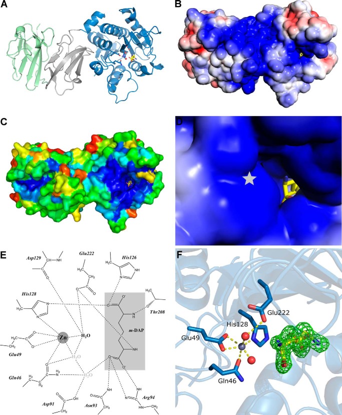
FIGURE 2.
The crystal structure of Csd4 (PDB code 4WCL). A, the overall monomeric structure of Csd4 with Zn2+ and m-DAP bound. The catalytic domain and domains 2 and 3 are colored blue, gray, and green, respectively. B and D, overall (B) and active site (D) magnified electrostatic surface potential of Csd4 contoured at ±3 kbT/ec. Electropositive regions are colored blue; electronegative regions are colored red; position of buried Zn2+ indicated with a star. C, distribution of conserved residues mapped onto the surface of Csd4. Most conserved regions are colored blue; the least conserved is colored red. E, two-dimensional interaction map between Csd4 and the product m-DAP (light gray). The predicted catalytic water highlighted in bold type. F, corresponding Zn-Csd4 active site with key ligands and an omit Fo − Fc difference density map for the density of the bound m-DAP product contoured at 3 σ.

