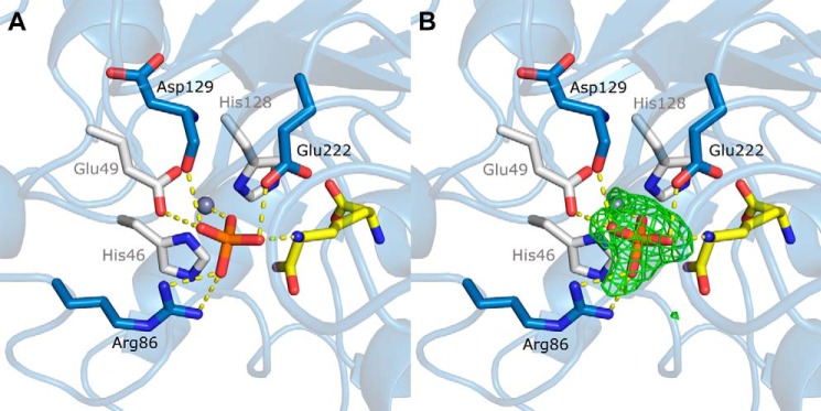
FIGURE 7.

Active site of the zinc-bound Q46H variant (PDB code 4WCM). A, zinc ligands are colored white; m-DAP is in yellow; phosphate is in orange; other residues interacting with the phosphate are colored blue; and phosphate interactions are shown as dotted lines. B, omit Fo − Fc difference density map contoured at 3 σ showing density for a bound phosphate.
