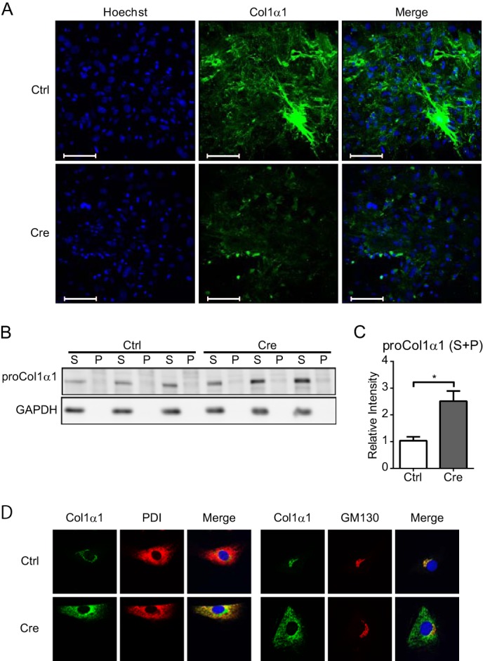
FIGURE 2.

Maturation of type I collagen in Hsp47-KO HSCs. A, deposition of type I collagen in the extracellular matrix after infection of activated HSCs with AdControl or AdCre at a MOI of 17 was detected by staining with an anti-type I collagen (green) antibody and Hoechst 33342 (blue) without permeabilizing cells. Scale bars, 100 μm. B, Western blot analyses of type I procollagen α1 in activated HSCs infected with AdControl or AdCre at a MOI of 25. Cell lysates were separated by centrifugation to generate detergent-soluble (S) and detergent-insoluble (P) fractions. C, the protein level of type I procollagen α1 in B is shown relative to that of GAPDH. Experiments were performed four times independently, and values are means ± S.E. *, p ≤ 0.05. D, the localization of type I collagen in activated HSCs infected with AdControl or AdCre at a MOI of 20 was determined by staining with anti-type I collagen (Col1α1; green), anti-protein-disulfide isomerase (PDI; left, red), and anti-GM130 (right, red) antibodies and Hoechst 33342 (blue). Ctrl, control.
