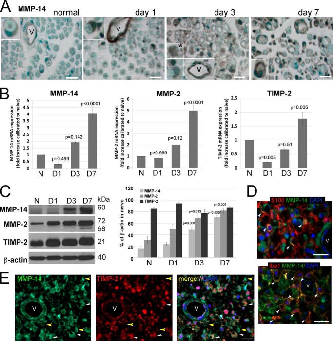
FIGURE 3.
The MMP-14/MMP-2/TIMP-2 axis in the PNS. A, immunostaining for MMP-14 using 3G4 antibody (2′2-diaminobenzidine; brown) in rat sciatic nerves at day 0 (normal) and days 1, 3, and 7 after crush injury (the crush site). MMP-14 is observed in Schwann cells (crescent-shaped; insets) and vessel (V) endothelial cells in all nerves and in macrophage-like cells at day 3 postinjury (asterisk). Images are representative of n = 3–4/group. Scale bars are 25 μm. B, TaqMan qPCR of MMP-14, MMP-2, and TIMP-2 in rat sciatic nerves at day 0 (normal (N)) and days 1, 3, and 7 after crush injury. The mean relative mRNA of n = 4/group are normalized to GAPDH and compared with the normal nerve samples (p values by analysis of variance and Bonferroni post hoc test) is shown. C, immunoblotting for MMP-14 (60 kDa), MMP-2 (72 and 68 kDa, latent and active, respectively), and TIMP-2 (21 kDa) in rat sciatic nerve at day 0 (normal) and days 1, 3, and 7 after crush injury. The graph represents the mean optical density of n = 4/group as a percentage of β-actin (p values by analysis of variance and Bonferroni post hoc test). D, immunostaining for MMP-14 (3G4 antibody; green) in the injured nerve with Schwann cells (S100; top, red) or macrophages (Iba1; bottom, red) depicts co-localization of the signals in the injured nerve (arrowhead). E, immunostaining of MMP-14 (3G4 antibody; green) and TIMP-2 (red) in rat sciatic nerve at day 3 postcrush. Schwann cells co-express MMP-14 and TIMP-2 (crescent-shaped structures; white arrows). TIMP-2−/MMP-14+ structures are observed (yellow arrowhead). D and E, all sections show DAPI-stained nuclei (blue) and vessels (V). Images are representative of n = 3/group. Scale bars are 25 μm. Error bars represent S.E.

