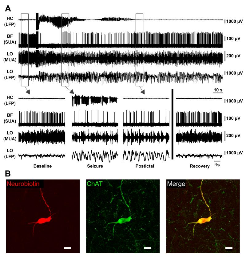
Figure 5.
Basal forebrain cholinergic neuron decreases firing during partial seizure.
(A) Cholinergic neuron in basal forebrain dramatically decreases firing during seizure activity. Expanded segments of baseline, seizure, and postictal recordings from the boxed regions in (A) as well as a recovery period 4.5 minutes after the stimulus.
(B) Labeled cholinergic cell recorded in (A) stained for Neurobiotin in left panel, choline acetyltransferase (ChAT) in middle panel, and merge in right panel. Scale bars are 20 micrometers. HC, hippocampus; BF, basal forebrain; LO, lateral orbitofrontal cortex; LFP, local field potential; SUA, single unit (juxtacellular) activity; MUA, multiunit activity. See also Figure S5

