Figure 8.
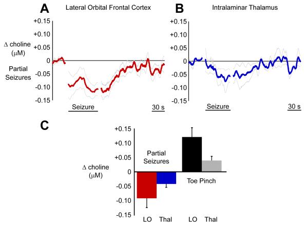
Choline signal decreases in the cortex and the thalamus during partial seizures.
(A) In the cortex, choline recordings decrease during partial seizures with gradual recovery during the postictal and recovery periods.
(B) The pattern is the same in the thalamus. Choline recordings decrease during partial seizures.
(C) Mean ictal changes in the cortex and the thalamus relative to 30 s baseline are shown along with mean changes during toe pinch to provide a common physiological change for comparison. Choline decreases in partial seizures were all statistically significant (paired t-tests Holm-Bonferroni corrected, P < 0.05). Toe pinch also elicited significant choline increases in cortex and thalamus (paired t-tests Holm-Bonferroni corrected, P < 0.05). Error bars in A-C are SEM. Mean timecourses in A and B are data 30 seconds prior to seizure onset, seizure timecourse scaled to mean seizure duration, and unscaled postictal timecourse aligned to seizure end. LO, lateral orbitofrontal cortex; Thal, intralaminar thalamus.

