
Figure 4.
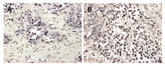
Histological examination of VX2 tumor in different groups. A: immunohistochemical stain showing the low expression of mutant-type p53 gene high in VX2 cells in group B, magnification × 400, B: The same stain showing the expression in group D, (× 400).