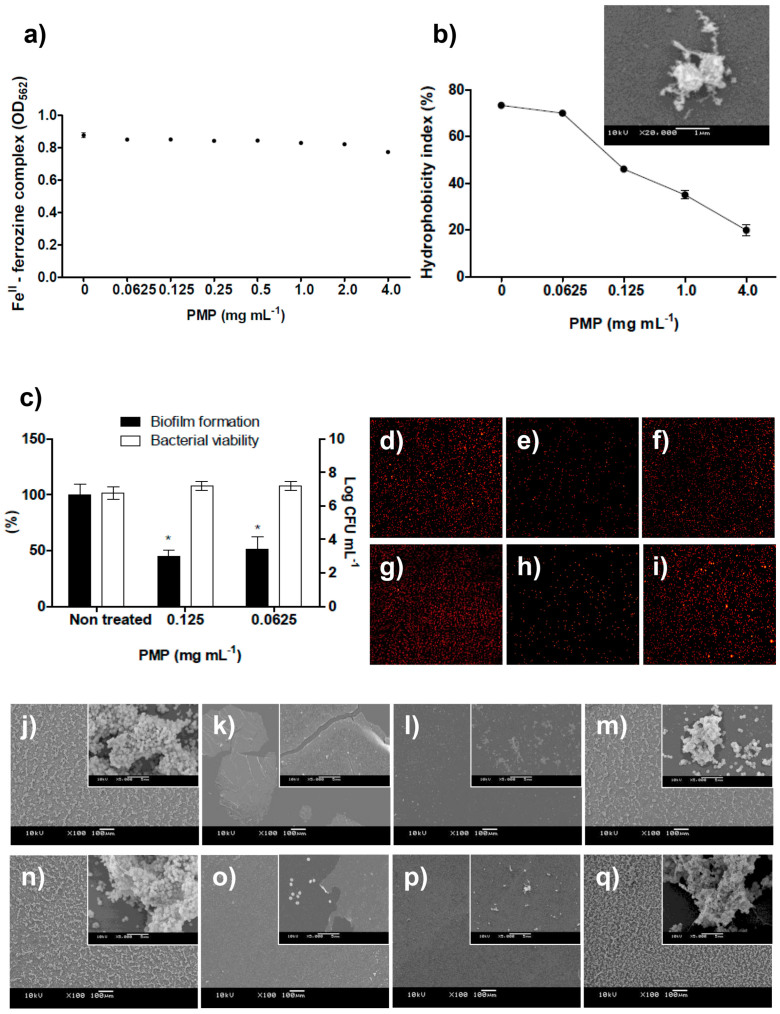
Figure 3.
(a) The concentration of ferrozine-FeII complex was not decreased in the presence of PMP, indicating that these proanthocyanidins are not strong FeII chelators (the standard curve established to determine the FeII concentration to be used in the ferrozine assay and the curve of positive-chelator 2,2-bipyridyl can be found in Supplementary Fig. S10). (b) The dose-dependent decreasing of S. epidermidis surface hydrophobicity index (HBPI); values of HPBI greater than 70% indicated hydrophobic bacterial surface (non-treated or treated with PMP at 0.0625 mg mL−1); less than 70% indicated hydrophilic bacterial surface. SEM image shows that surface of S. epidermidis cells become covered by amorphous material in solutions containing PMP. (c) S. epidermidis partially recovered the ability to form biofilm and remained viable after exposure to proanthocyanidins for 24 h with three subsequent washes using sterile 0.9% NaCl. * represents statistical difference (p < 0.05) between treated and non-treated samples when analyzed by Student's t-test. (d–i) Fluorescence microscopy images (10x magnification) demonstrated that (d and g) non-treated microspheres and (f and i) microspheres treated with 0.0625 mg mL−1 of PMP attach to Permanox and to glass surfaces, respectively, while attachment was inhibited for spheres exposed to 0.125 mg mL−1 of PMP (e and h, respectively for Permanox and glass surfaces), similarly as observed for S. epidermidis cells treated with PMP. similarly as observed for S. epidermidis cells treated with PMP. (j–q) SEM images of S. epidermidis treated with PMP. Untreated S. epidermidis displayed several microcolonies on (j) Permanox and (n) on glass surfaces. When S. epidermidis was exposed to 4.0 mg mL−1 of PMP, there was no sign of attached cells while some regions of (k) Permanox and (o) glass substrates, and PMP spontaneously adhered on hydrophobic and hydrophilic substrates (note the insert showing that single cells adhered where there is no PMP film). Inhibition of biofilm formation at 0.125 mg mL−1 of PMP is observed on (l) Permanox and on (p) glass surfaces. The ability of S. epidermidis to form protective biofilm is not inhibited by 0.0625 mg mL−1 of PMP neither on (m) Permanox nor on (q) glass substrates. Bars represent 100 μm and in the inserts, 5 μm. All of these methodologies can be found in Supplementary Methods.

