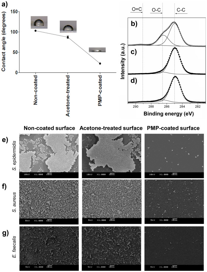
Figure 4. Green-coated surface analyses.
(a) WCA of non-coated, acetone-treated and PMP coated-surfaces. (b) XPS analyses of PMP coated-surfaces, (c) acetone-treated and (d) non-coated surfaces. SEM images of adhesion and biofilm formation by (e) S. epidermidis, (f) S. aureus and (g) E. faecalis on non-coated surface, acetone-treated surface and PMP-coated surface, respectively. Bars indicate 10 μm.

