Figure 1.

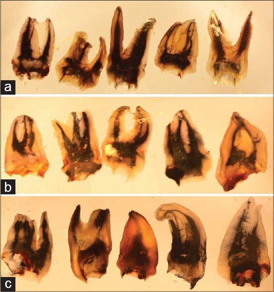
Photographs of injected dye and cleared specimens; maxillary first molars (a); second molars (b) and third molars (c) showing various roots with root canal numbers and canal anatomies

Photographs of injected dye and cleared specimens; maxillary first molars (a); second molars (b) and third molars (c) showing various roots with root canal numbers and canal anatomies