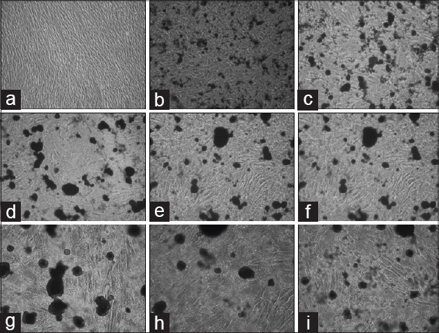
Figure 3.

Image of periodontal ligament (PDL) cells after application of 2% Chlorhexidine gluconate (CHX) with an inverted microscope. Both numerical reduction and morphological differences have been identified in PDL cells. Fusiform morphology of PDL cells has undergone differentiation. (a) Control group; human periodontal ligament (hPDL) Inverted Microscope Image (IMG ×10) that taken from unapplied CHX (b-e) hPDL image (IMG ×10) that taken 1 h intervals after CHX application. (f-i) hPDL image (IMG ×10) that taken 1 h intervals after 24 h CHX application
