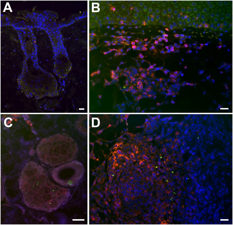
FIGURE 4.
Early subepidermal infiltrates in eschar biopsies of cynomolgus macaques. Post-ID inoculation O. tsutsugamushi localize to the hair follicles and sebaceous glands (A and C) accompanied by formation of early CD3+ lymphocyte infiltrates. The subdermal infiltrates in cynomolgus macaques at day 7 postinoculation of O. tsutsugamushi are characterized by a large proportion of T lymphocytes (B), as well as formation of lymphocyte follicles in the deeper dermis (D), with localization of O. tsutsugamushi at the peripheral border, similar to the location also seen in spleen follicles (data not shown). Double immunolabeling with CD3 staining is in red and O. tsutsugamushi is in green. (A) NHP ID6856, day 2 biopsy. (B and D) NHP ID5234, day 7 biopsy. (C) NHP ID5234, day 2 biopsy. All images were taken using a Nikon Eclipse E400 microscope and Nikon digital camera (DS-L1; Nikon, Tokyo, Japan); images were merged and optimized with Photoshop CS3 extended, version 10.0. Original magnification ×100 (A); ×400 (B and D); ×600 (C). Scale bars, 20 μm.

