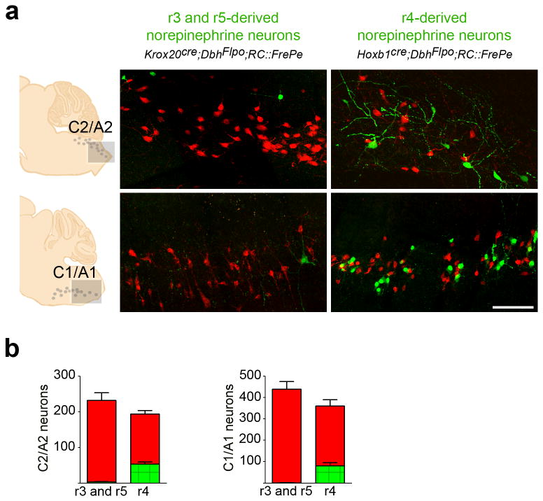
Fig. 3. r3&5(Krox20cre)- and r4(Hoxb1cre)-derived norepinephrine neurons populate the medullary C1/A1 and C2/A2 brainstem nuclei.
(a) Sagittal sections from adult mouse brainstems reveal the contribution of norepinephrine neurons derived from r3&5(Krox20cre;DbhFlpo;RC∷FrePe) and r4(Hoxb1cre;DbhFlpo;RC∷FrePe) to the caudal regions of the C1/A1 and C2/A2 nuclei. Our analyses do not distinguish epinephrine neurons in C2 and C1 from norepinephrine neurons in A2 and A1. eGFP (green) marks the r-derived population and mCherry (red) marks all other norepinephrine and epinephrine neurons in the representative sections corresponding to the boxed areas within the schematics (left). Scale bar indicates 100 μm. (b) Cell counts of r-derived norepinephrine and epinephrine neurons in the C1/A1 and C2/A2 medullary nuclei (r3&5 n=5; r4 n=6 mice; error bars are mean ± s.e.m.). Unpaired, two-tailed t-tests demonstrate that total numbers of norepinephrine and epinephrine neurons (sum of eGFP- and mCherry-positive cells) were not significantly different between genotypes (p>0.05; df=9; C2/A2 t=1.571; C1/A1 t=1.631).

