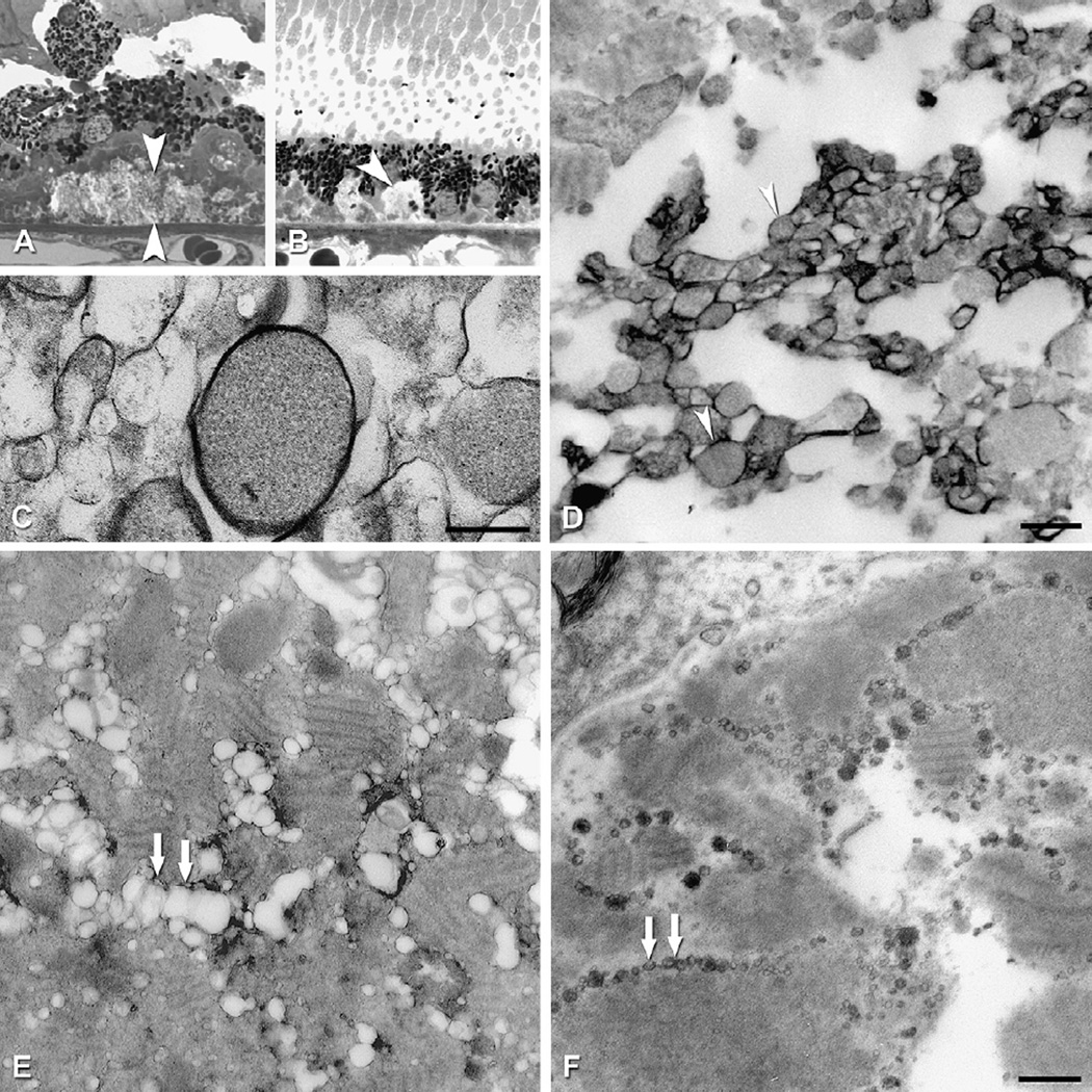
Fig. 14.
Ultrastructure of cholesterol-containing components of drusen and deposits. Tissue post-fixed with OTAP, from (Curcio et al., 2005b). A. RPE migrating anteriorly and thick BlamD containing cellular processes, bracketed by large arrowheads. B. BlamD containing basal mounds of membranous debris (arrowhead). Bar, 200 nm C. Membranous debris with contents, interior of a large druse. Bar, 200 nm. D. Individual profiles in the basal mound of panel B are solid and surrounded by an electron-dense band. E,F. Bar is 500 nm. E. Linear tracks of partially extracted material resembling membranous debris (double arrows) in BlamD. F. Linear tracks of solid particles (double arrows) in BlamD of the same eye as E, post-fixed with OTAP.

