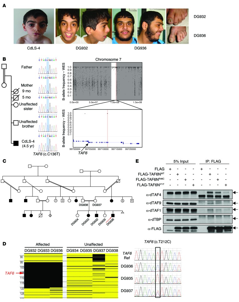
Figure 5. TAF6 variants identified in the Turkish family of CdLS-4 and the Saudi family.
(A) Patient photographs showing the clinical features of CdLS-4 and 2 patients (DG932 and DG936) from the Saudi family. (B) The pedigree of the CdLS-4 family and the segregation of the TAF6 variant. B-allele frequency plots from the WES data for CdLS-4 are shown on the right. The region of AOH is shown as a white region between the flanking gray areas. Top panel: B-allele frequency plot of the entire chromosome 7; bottom panel: zoomed-in view of the region encompassing TAF6 indicated by the black arrow. (C) The pedigree of the Saudi family. Red arrow indicates the patient that underwent WES. (D) The homozygosity mapping on the left shows the region of AOH (black blocks) encompassing TAF6 (indicated by red arrow) in DG932, DG933 and DG936. The Sanger-sequencing chromatograms on the right show homozygous WT in the control and DG935, homozygous mutation in DG936, and heterozygous mutation in the mother, DG937. (E) Co-IP in Drosophila S2 cells testing TAF6-binding affinity. S2 cells were treated with dsRNA targeting dTAF6C. FLAG-TAF6NWT, FLAG-TAF6NR46C, and FLAG-TAF6NI71T correspond to the S2 cells transfected with WT or mutated dTAF6N as indicated. FLAG, negative control. Four TFIID components (dTAF1, dTAF4, dTAF9, and dTBP) were detected with Western blot using antibodies against each indicated component. Left 4 columns, input; right 4 columns, proteins co-IP with FLAG-TAF6N. The lanes were run on the same gel, but were noncontiguous.

