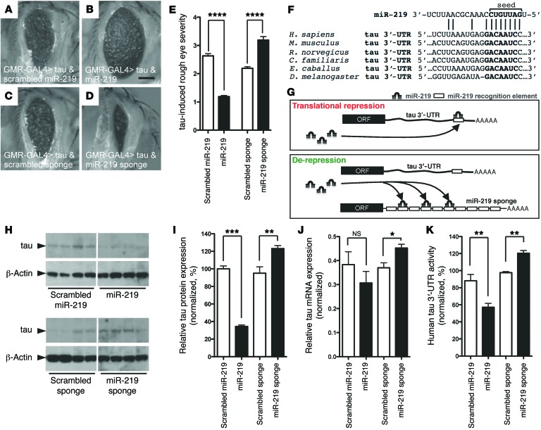
Figure 2. In vivo regulation of tau toxicity and expression by miR-219.
(A and B) Coexpression of miR-219 partially suppressed the human tau–induced rough eye phenotype compared with that seen in the scrambled miR-219. (C and D) Coexpression of the miR-219 sponge with human tau exacerbated the phenotype compared with that observed in the scrambled sponge. Scale bar: 100 μm. (E) Semiquantitative assessments of the rough eye phenotype (n = 38/group). (F) miR-219 recognition element in the tau 3′-UTR. (G) Schematic illustrating regulation of tau by miR-219 and de-repression with the sponge. (H and I) Quantitative immunoblot of extracts from flies expressing miR-219 in the brain revealed decreased Drosophila tau protein levels compared with those detected in the scrambled control. Expression of the miR-219 sponge shows increased tau protein levels compared with those in the scrambled control (noncontiguous from the same gel). (J) qPCR shows that the tau mRNA levels were unchanged in the miR-219–expressing flies, but there was an increase in tau mRNA levels in flies expressing the miR-219 sponge compared with that observed in the scrambled control. (K) Flies that coexpress miR-219 and luciferase fused to the human tau 3′-UTR showed reduced activity compared with that seen in the scrambled control. Conversely, the miR-219 sponge significantly increased 3′-UTR activity compared with that in the scrambled control. Flies expressing a GAPDH 3′-UTR fused to luciferase were used for normalization. Data are representative of 3 experiments. *P ≤ 0.05, **P ≤ 0.01, and ***P ≤ 0.001 by 2-tailed Student’s t test; ****P ≤ 0.0001 by Mann-Whitney U test.

