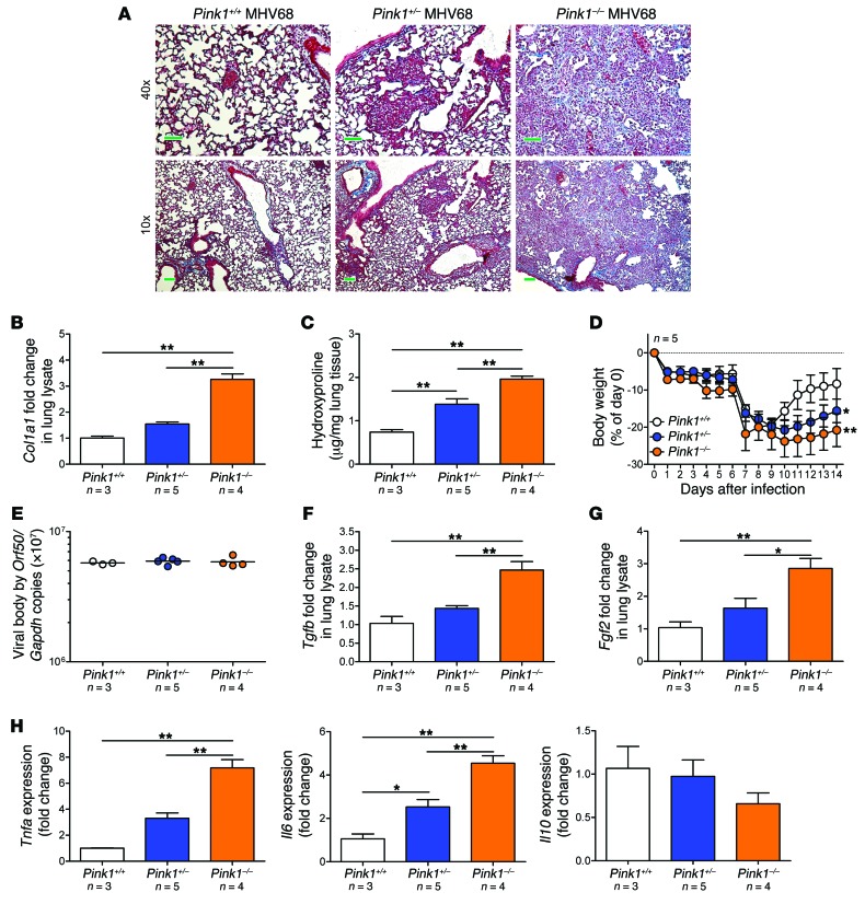
Figure 11. PINK1 deficiency increases susceptibility to lung fibrosis.
(A) Representative Masson trichrome staining in Pink1+/+, Pink1+/–, and Pink1–/– lung sections showed increased collagen deposition (blue) at day 15 after MHV68 infection. Scale bars: 10 μm (40×); 50 μm (10×). (B) Higher Col1a1 transcript levels in lungs of PINK1-deficient mice infected with MHV68 compared with control littermates. (C) Increased collagen deposition (assessed by hydroxyproline level) in lungs of PINK1-deficient mice after infection. (D) Weight loss after MHV68 infection was more severe in Pink1–/– mice. (E) Viral load (assessed by qPCR) of individual mouse lungs 15 days after MHV68 infection. Bars represent geometric mean. (F and G) Relative change in lung lysate Tgfb (F) and Fgf2 (G) transcript levels after infection. (H) Relative change in Tnfa, Il6, and Il10 mRNA levels after infection. Data represent mean ± SEM (B–D and F–H). *P < 0.05, **P < 0.01, 1-way ANOVA (B, C, and E–H) or 1-way repeated-measures ANOVA (D) with post-hoc Bonferroni.

