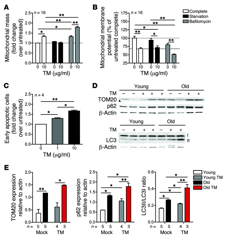
Figure 5. Stimulation of ER stress deteriorates mitochondrial function and impairs mitophagy in lung epithelial cells.
(A) A549 cells were treated with or without TM (1 μg/ml for 24 hours), and mitochondrial mass was determined by MitoTracker Green. Induction of autophagy by serum starvation reduced mitochondrial mass in TM-treated cells. The autophagy inhibitor bafilomycin A1 increased mitochondrial mass in untreated and TM-treated cells. (B) TM induced dose-dependent depolarization of mitochondria in A549 cells (assessed by JC-1 dye staining). Depolarization was increased in the presence of bafilomycin A1, but was not affected by starvation conditions. (C) Increased doses of TM induce apoptosis of A549 cells (assessed by annexin V staining). (D) Representative Western blot analyses showing increased levels of the mitochondrial marker TOM20 and autophagy markers p62 and LC3I/LC3II in lung lysates from aging and young mice after vehicle and TM treatment (2 μg/mouse). The β-actin blot was obtained from parallel samples run on a separate gel from the TOM20 and p62 blots. (E) Density analyses of Western blots in D. Data represent mean ± SEM (A–C and E). *P < 0.05, **P < 0.01, 1- (A–C) or 2-way (E) ANOVA with post-hoc Bonferroni.

