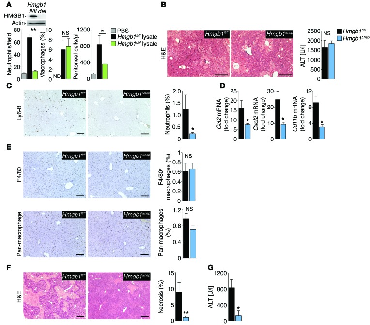
Figure 2. HMGB1 promotes neutrophil recruitment in vitro and in vivo.
(A) Neutrophil and macrophage migration toward Hmgb1-floxed and Hmgb1-deleted (Hmgb1del) liver extracts (induced by Mx1-Cre), determined in Boyden chambers (left 2 panels, n = 3 per group, with representative results from 3 separate isolations). Insert shows immunoblot confirming Hmgb1 deletion. Peritoneal inflammatory cell accumulation after i.p. injection of lysates from Hmgb1fl/fl (n = 7) and Hmgb1del (n = 8) livers. (B–E) Hmgb1fl/fl (n = 8) and Hmgb1Δhep (n = 9) mice were subjected to warm hepatic I/R and sacrificed 6 hours later. H&E staining (B, left panel) and serum ALT (B, right panel) demonstrate similar initial injury, whereas hepatic neutrophil infiltration (C) and hepatic expression of inflammatory genes (D) differed. Numbers of hepatic macrophages determined by F4/80 staining and staining with pan-macrophage antibody in Hmgb1fl/fl (n = 8) and Hmgb1Δhep (n = 9) mice (E). (F and G) Hepatic injury 24 hours after I/R injury in Hmgb1fl/fl (n = 11) and Hmgb1Δhep (n = 10) mice, determined by H&E staining (F) and serum ALT (G). *P < 0.05 and **P < 0.01 by 1-way ANOVA followed by Tukey’s multiple comparisons test (A) and unpaired 2-tailed t test (B–G). Scale bars: 200 μm.

