
Figure 3. Immunohistochemical localization of PC in lung tissues.
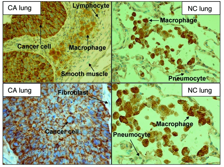
Pairs of CA and NC tissues obtained from NSCLC patients were stained with anti-PC antibody. Shown are representative pairs of micrographs from 1 patient. Original magnification, ×400 (top panels) and ×600 (bottom panels). Additional patient data are provided in Supplemental Figure 3. In CA tissues, viable cancer cells stained intensely for PC, while macrophages stained weakly, and other stromal cells showed no staining for PC. In adjacent NC tissues, macrophages stained strongly for PC, while pneumocytes stained very weakly for the protein.