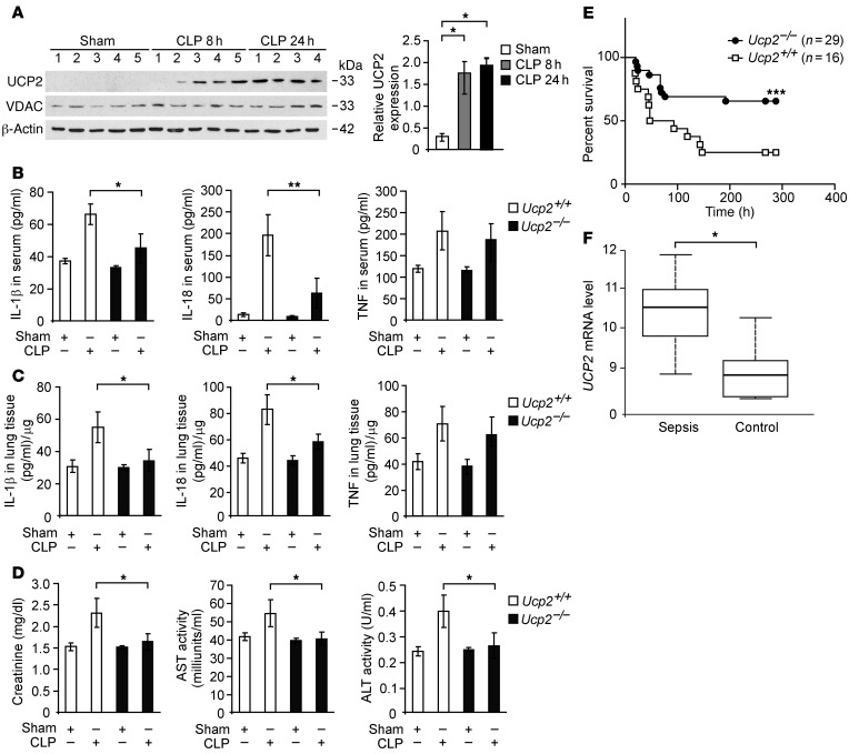
Figure 1. Deficiency of UCP2 protects against CLP-induced mortality in vivo.
(A) Immunoblot analysis of UCP2 and voltage-dependent anion channel (VDAC) in lung tissues from wild-type mice 8 and 24 hours after CLP or sham laparotomy. β-Actin served as the standard. *P < 0.05, ANOVA. (B) ELISA assay for IL-1β, IL-18, and TNF in the sera and (C) in the lung tissues from Ucp2+/+ or Ucp2–/– mice 24 hours after CLP or sham laparotomy. For both Ucp2+/+ and Ucp2–/– mice, sham, n = 2, and CLP, n = 8. *P < 0.05, **P < 0.01, ANOVA. (D) Organ dysfunction of CLP was determined in Ucp2+/+ or Ucp2–/– mice 24 hours after CLP or sham laparotomy. Organ dysfunction biomarkers, including creatinine, aspartate aminotransferase (AST), and alanine aminotransferase (ALT), were measured in sera. For both Ucp2+/+ and Ucp2–/– mice, sham, n = 2, and CLP, n = 8. *P < 0.05, ANOVA. (E) Survival curve of CLP was determined in Ucp2+/+ or Ucp2–/– mice. Ucp2+/+, n = 16; Ucp2–/–, n = 29. ***P < 0.0055, log-rank test. (F) Box plots comparing measures of UCP2 mRNA levels in whole-blood samples of sepsis (n = 30) and control (n = 19) patients. The UCP2 mRNA levels are presented as median value (black line), interquartile range (box), and minimum and maximum of all data (whiskers). *P < 0.05, ANOVA.

