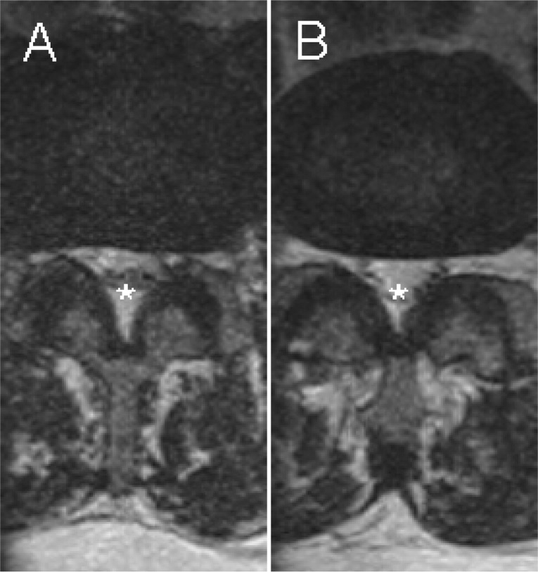
Figure 4.
T2-weighted coronal spot view MR images of L3–4 (A) and L4–5 (B) showing disc protrusion, bilateral facet joint osteoarthritis, and ligamentum flavum hypertrophy, with resultant central canal (asterisks), lateral recess, and intervertebral foraminal stenosis (most severe at L3–4), bilaterally.

