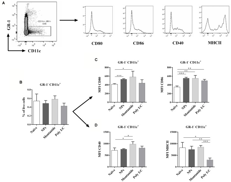
FIGURE 4.
Dendritic cell activation in dLNs after injection with PSNPs, Montanide, and Poly I:C. Mice (BALB/c) were injected once intradermally at the base of tail with the different adjuvants alone. 48 h after injection, mice were sacrificed, local (inguinal) dLNs were harvested and the levels of CD11c+ DCs and various activation markers were assessed by flow cytometry. (A) gating strategy; (B) frequency of GR-1-CD11c+ cells; (C) Mean fluorescent intensity (MFI) of CD80 and CD86 on GR-1-CD11c+ cells; (D) MFI of CD40 and MHCII on GR-1-CD11c+ cells. Data presented as mean ± SD of MFI for each group of treatment (n = 3 mice/group). Statistical analysis was performed via t-tests, *p ≤ 0.05, **p ≤ 0.01, ***p ≤ 0.001.

