
Figure 4.
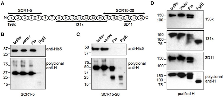
Mapping of cleavage sites in factor H. (A) Graphic of factor H indicating the areas covered by the recombinant fragments SCR1-5 and SCR15-20 and the localization of the 196X, 131X, and 3D11 mAb epitopes. (B–D) E. coli 83972 ΔompT bacteria expressing PgtE, Pla or the empty vector were incubated for 1 h with equimolar amounts of recombinant H fragments SCR1-5, SCR15-20 or purified H. The non-reduced supernatants were run in SDS-PAGE and immunoblotted. (B) SCR1-5 and (C) SCR15-20 blotted with a monoclonal anti-His5 Ab recognizing the C-terminal His8-tag in the H fragments, and with a polyclonal anti-H Ab. (D) Purified H blotted with monoclonal Abs 196X, 131X, and 3D11 recognizing the N-terminal SCR1, the SCRs 8-14.5 and the SCR18, respectively; or with a polyclonal anti-H Ab. The assays were conducted at least twice with consistent results.