
Figure 6.
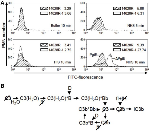
The effect of PgtE on opsonophagocytosis of S. enterica. (A) FITC-stained S. enterica 14028R and 14028R-1 (ΔpgtE derivative of 14028R) were incubated for 5 min in 50% NHS, 50% HIS or buffer. Freshly isolated human neutrophils were mixed with the bacteria at a MOI 20, and phagocytosis was allowed to take place for 5 min (in NHS) or 10 min (in NHS, HIS or buffer). The FITC signals from 10,000 paraformaldehyde-fixed neutrophils were measured using flow cytometry. Median values of fluorescence are shown. Striped area, 14028R; open area, 14028R-1. The experiment was conducted three times, each time with PMN from a different donor and with consistent results. A representative result is shown. (B) Progression of the alternative complement pathway up to the level of iC3b that is the main opsonic ligand for CR3 (CD11b/CD18) on neutrophils. Arrowheads indicate proteolytic cleavage. Slashes mark the components cleaved by PgtE.