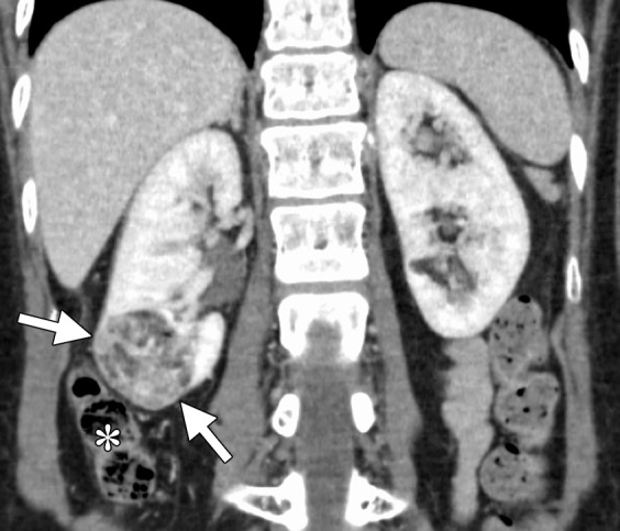
Figure 10a.

CT images in a 55-year-old woman with a history of right renal angiomyolipoma with associated massive retroperitoneal hemorrhage that required immediate embolization. (a) Coronal reconstructed image from follow-up contrast-enhanced CT shows continued avid enhancement of the lesion (arrows). Note the closely adjacent colon (*). After successful hydrodissection for displacement of the colon, the lesion was targeted for microwave ablation with three 17-gauge gas-cooled antennae at 65 W for 5 minutes. (b) Coronal reconstructed image from contrast-enhanced CT 4 months after ablation shows minimal, if any, residual enhancement of the lesion (arrows). The patient has remained asymptomatic.
