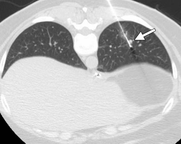
Figure 12a.

CT images in a 54-year-old woman with a history of metastatic colorectal cancer. (a) Axial CT image shows a 6-mm biopsy-proven metastatic focus in the left lower lobe of the lung (arrow) that was targeted for RF ablation. A single 17-gauge water-cooled electrode was positioned in the medial aspect of the nodule, and a 12-minute ablation was performed. (b) Axial CT image obtained 6 months after ablation shows rapid local tumor progression, with the metastatic focus now measuring 1.6 cm. This area was targeted for repeat RF ablation by using a water-cooled cluster electrode. The repeat ablation resulted in long-term local control but was complicated by a large hemothorax that ultimately required surgical intervention. The hemothorax was at least partially related to the increased invasiveness of the cluster electrode.
