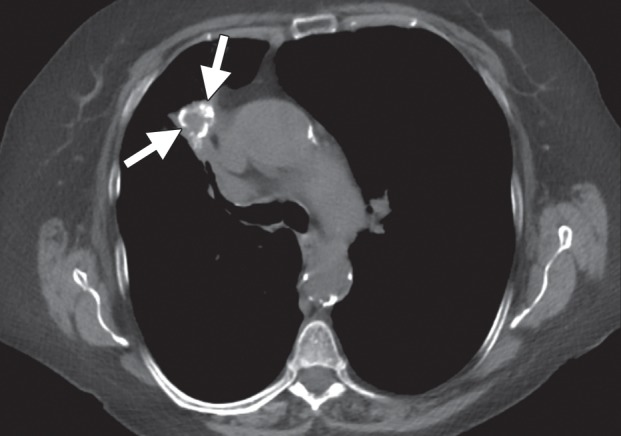
Figure 13c.

Imaging findings in a 71-year-old woman with a history of non–small-cell lung carcinoma who had undergone radiation therapy 2 years previously. (a) Axial positron emission tomography (PET)/CT image shows recurrent disease in the radiation field (arrows). The patient was referred for ablation. Cryoablation was chosen because of the close proximity of the mass to the mediastinum and the associated risk for injury to the phrenic nerve. (b) Axial CT image shows the four cryoprobes (arrow) used to perform the ablation, which involved a 3-, 5-, 7-, 5-, 5-minute freeze-thaw protocol. This resulted in excellent coverage with the ice ball (arrowhead). Phrenic nerve palsy developed, with associated diaphragmatic paralysis. Combined with poor baseline lung function, this complication resulted in a prolonged postablation hospitalization. Within 5 weeks, however, both diaphragmatic function and respiratory status returned to baseline levels. (c) Axial nonenhanced CT image obtained 18 months after ablation shows calcification of the treated mass (arrows), with no evidence of local or distant recurrence.
