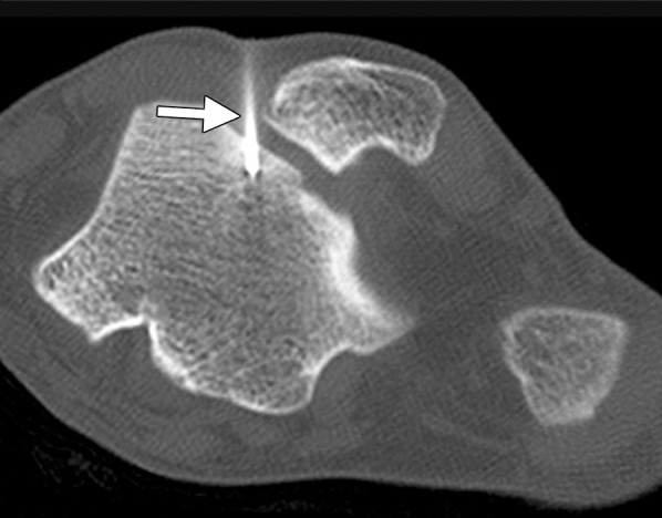
Figure 18b.

CT images in a 16-year-old male patient with lateral ankle pain. (a) Coronal nonenhanced CT image shows an osteoid osteoma of the lateral talar dome (arrow). (b) Because a surgical approach would be associated with substantial potential morbidity, the osteoid osteoma was targeted for treatment with RF ablation. As shown on an axial CT image, a single 17-gauge electrode with a 1-cm active tip (arrow) was placed in the lesion with CT fluoroscopic guidance, and a 6-minute ablation was performed. The symptoms completely resolved, and the patient remains asymptomatic. (Case courtesy of Kirkland Davis, MD.)
