
Figure 3a.
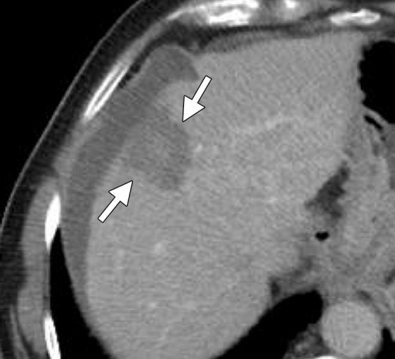
Computed tomographic (CT) images in a 77-year-old man with cirrhosis and a 2.6-cm hepatocellular carcinoma (HCC) in the right hepatic lobe. The HCC was targeted for microwave ablation with a single, specially designed “precision” 17-gauge gas-cooled antenna at 65 W for 7.5 minutes. The resulting ablation zone (arrows) shown on axial (a) and coronal (b) contrast material–enhanced postablation CT images is round and measures approximately 3.4 cm in all dimensions. Microwave ablation allowed destruction of the HCC with minimal associated damage to the hepatic parenchyma. Note the artificial ascites used to protect the diaphragm (arrowhead in b).