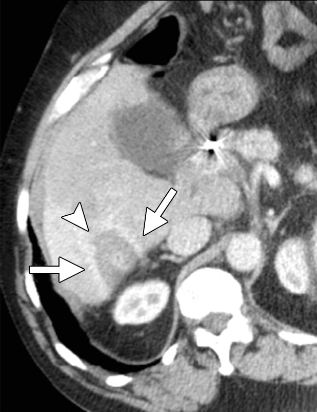
Figure 4c.

CT images in a 64-year-old male patient with cirrhosis and HCC. (a) Axial arterial phase biphasic CT image shows a 1.8-cm HCC in the right hepatic lobe (arrows). This was targeted for microwave ablation with a single 17-gauge gas-cooled antenna. A 3-minute ablation was performed at 90 W. (b) Axial nonenhanced CT image obtained during ablation shows that the mass has essentially been replaced by gas (arrow). (c) Axial CT image obtained immediately after ablation shows a 3.0 × 2.8-cm ablation zone (arrows), with complete ablation of the tumor and only the expected benign periablational enhancement (arrowhead). The high attenuation seen centrally in the ablation zone is desiccated dense tissue, which is more prominent with microwave ablation than with RF ablation.
