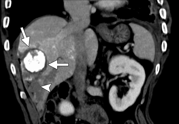
Figure 5b.

CT images in a 55-year-old male patient with cirrhosis and HCC. (a) Axial nonenhanced CT image obtained during transarterial chemoembolization shows a 5.4 × 5.2-cm HCC in the right hepatic lobe, with intense uptake of ethiodized oil (arrows) and less intense nontarget uptake in the remainder of the liver. The tumor was targeted for microwave ablation by using US guidance. Three 17-gauge gas-cooled microwave antennae were positioned in a triangular configuration within the tumor, and 65 W was applied to all antennae simultaneously, for a total ablation time of 10 minutes. (b) Sagittal reconstructed contrast-enhanced CT image obtained immediately after ablation shows that the ablation was technically successful, with an adequate circumferential ablative margin (arrows). Note the necrosis in the nontarget liver related to transarterial chemoembolization (arrowhead).
