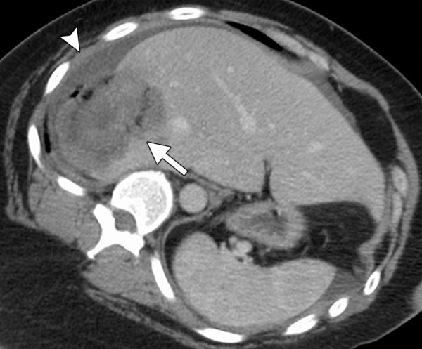
Figure 6c.

CT images in a symptomatic 37-year-old woman. (a) Axial contrast-enhanced portal venous phase CT image shows an 8.8 × 8.7-cm giant hemangioma in the right hepatic lobe (arrows). (b) Sagittal unenhanced CT image shows the hemangioma targeted for microwave ablation, with three 17-gauge gas-cooled microwave antennae distributed evenly in the hemangioma (arrows). (c) Axial contrast-enhanced CT image obtained after 15 minutes of ablation at 65 W shows complete ablation of the tumor, with a small margin of normal hepatic tissue (arrow). Note the hydrodissection fluid (arrowhead), which was used to protect the abdominal wall and diaphragm.
