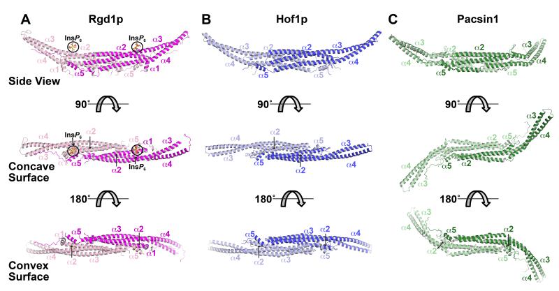
Figure 3. Structures of the Rgd1p and Hof1p F-BAR domains.
(A) Cartoon of the dimeric Rgd1p F-BAR domain, with one molecule colored magenta (right) and the other pink (left). Three views are shown, with the F-BAR domain progressively rotated around its long axis. The middle (orthogonal) view looks into the concave surface, and the bottom view is 180° rotated so that the view is into the convex surface. The five primary α-helices are labeled. The two bound InsP6 molecules (bound to the concave surface) are labeled and circled in black. See also Figures S2 and S3.
(B) The Hof1p F-BAR domain represented as in (A), but with the two monomers colored dark blue (right) and light blue (left). See also Figures S2 and S3.
(C) Structure of the Pacsin-1 F-BAR domain from PDB entry 3HAI (Wang et al., 2009), illustrating the ‘S’ or tilde shape seen in some F-BAR domains.

