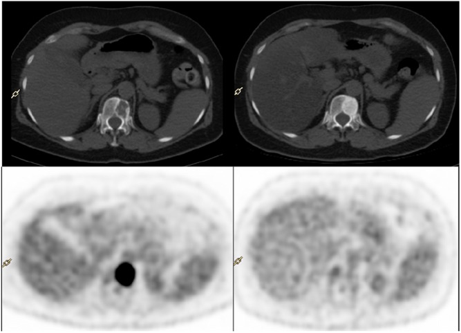
Figure 1.
Example of fluorodeoxyglucose-positron emission tomography (PET)/computed tomography (CT) exam before (left) and after (right) 3 months of eribulin and trastuzumab therapy in a woman with an osteolytic metastases of breast cancer, located on TH12 . After treatment, CT showed an important sclerotic reaction of the bone lesion, whereas PET showed no more metabolic activity. Both the anatomic and molecular imaging are in favor of a good response to systemic treatment.

