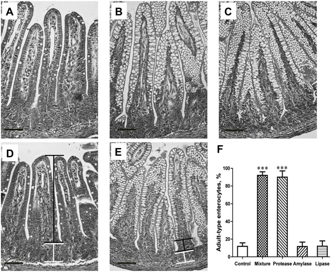
Fig 3. Photomicrographs of H&E stained distal small intestine sections from 17 d-old rats, showing the appearance of mature enterocytes, lacking supranuclear vacuoles, in the villi epithelium after gavage feeding with different microbial-derived enzymes, a protease (A), amylase (B), lipase (C), and a mixture thereof (D) or water (control, E) during 14–16 days of age in suckling rats.
The morphometric evaluation of the epithelial maturity, i.e., the portion of adult-type enterocytes, lacking supranuclear vacuoles, appearing on the villi is shown (F). The horizontal bar inserted indicates 100 μm; black connector line shows the portion of villus with adult-type epithelium, and white line shows the crypt region. Statistically significant differences between control and enzyme-treated groups (mean ± SD, n = 7) are indicated by *** P < 0.001.

