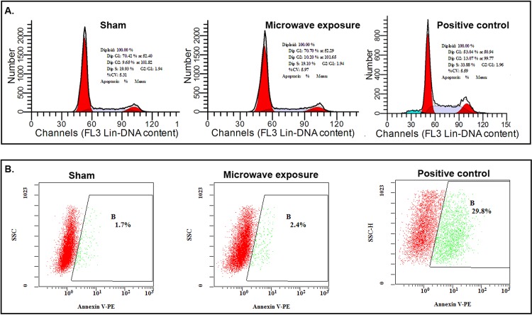
Fig 4. Cell cycle and apoptosis of MSCs detected with flow cytometry after 2.856 GHz microwave exposure at SAR of 4 W/kg.
(A) Cell cycle detected with PI. The representative results of sham, microwave exposure and the positive control were shown in the upper panel. (B) Apoptosis detected with annexin V-PE. The representative results of sham, microwave exposure and the positive control were shown in the lower panel.

