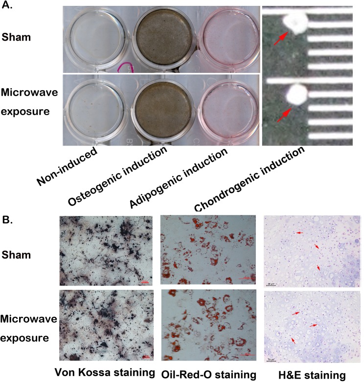
Fig 5. MSCs in vitro differentiation after 2.856 GHz microwave exposure at SAR of 4 W/kg.
(A) Osteogenic induction and adipogenic induction were detected with Von Kossa and Oil-Red-O staining. The induced chondrogenic pellets were placed onto a black ruler as indicated by the arrows. (B) Microscopic examination of induced osteoplasts (left panel), and adipocyted (middle panel) with Von Kossa and Oil-Red-O staining (scale bar = 100 μm), and chondrocytes (right panel) with H&E staining (scale bar = 50 μm).

