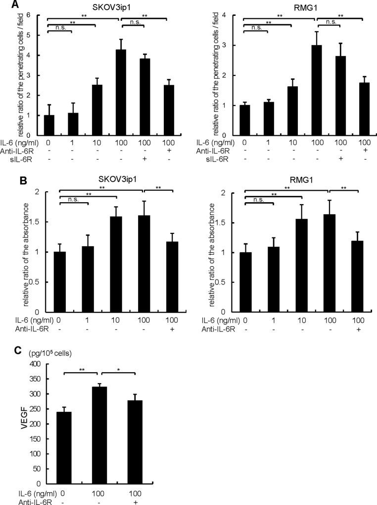
Fig 3. Exogenous treatment of IL-6 promotes ovarian cancer cell proliferation, invasion and VEGF production.
(A) A matrigel invasion assay was done using a modified Boyden chamber system. 1 x 10⁵ of SKOV3ip1 (left) or RMUS-S (right) cells were placed on the top chamber in serum-free medium and allowed to invade for 72 h. Various concentrations of IL-6 (1–100 ng/ml) or 60 ng/ml of sIL-6R were applied in the bottom chamber as a chemoattractant. 10 μg/ml of anti-IL-6R antibody or non-immune IgG was co-treated. Non-invading cells were removed using a cotton swab, and invading cells on the underside of the filter were enumerated. Relative numbers of invading cells with respect to the control (no IL-6 treatment) are shown. (B) In vitro cell proliferation assay. 1 x 10⁴ cells of SKOV3ip1 (left) or RMG1 (right) cells were plated in 24-well plates in 10% FBS/DMEM for 24 h and then incubated in serum-free medium in the presence or absence of various concentrations of IL-6 (1–100 ng/ml) with or without anti-IL-6R antibody or non-immune IgG as control for 72 h. Cell proliferation was evaluated by a modified MTS assay. Cell proliferation was expressed as the ratio of the number of viable cells. (C) ELISA assay of VEGF-A. 1 x 10⁵ SKOV3ip1 cells were plated onto 6-well plates and cultured with 2 ml of serum-free medium in the presence or absence of 100 ng/ml of IL-6 for 72 h. Anti-IL-6R antibody or control IgG was co-treated. Conditioned media were collected and the concentration of human VEGF-A was measured by ELISA. Experiments were repeated three times and values are means ± SD of triplicates. n.s.; not significant, *; P < 0.05, **; P < 0.01.

