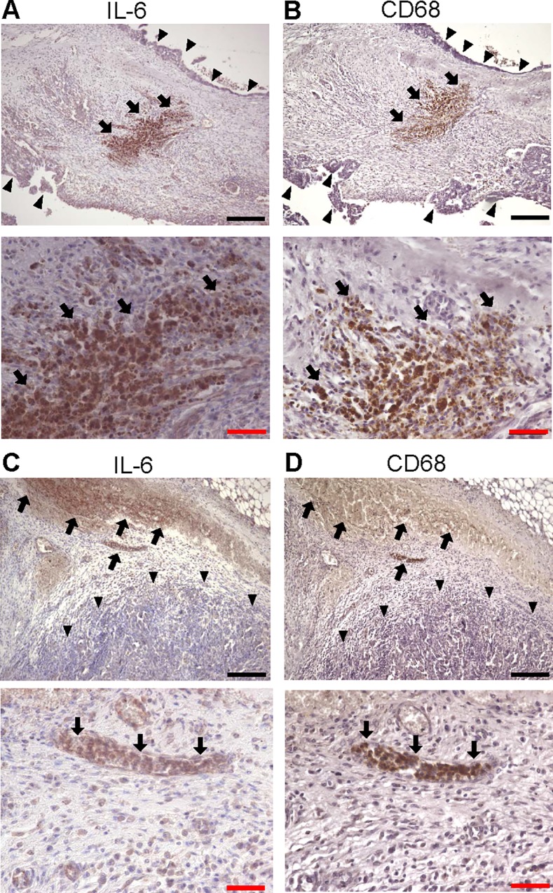
Fig 4. Immunohistochemical analyses of IL-6 in high-grade serous ovarian cancer tissues.
Serial sections of stage III high-grade serous ovarian cancer tissues were immunostained with anti-IL-6 antibody (A, C) and anti-CD-68 antibody (B, D). IL-6 was strongly expressed in stroma, while cancer cells little expressed IL-6. (A, B) Sections from a 56 year-old female with stage IIIC high-grade serous ovarian cancer. (C, D) sections from a 63 year-old female with stage IIIC high-grade serous ovarian cancer. CD68 staining identified macrophages. Arrows indicate macrophages. Arrowheads indicate ovarian cancer cells. Original magnification, x100 (upper panels), and x400 (bottom panels). Black bar; 200 μm, red bar; 50 μm.

